
What is Pterygium?
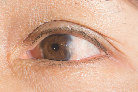
Pterygium, also known as surfers’ eye, is a benign growth of the conjunctiva (a layer over the white part of the eye) that grows into the cornea (the clear layer over the colored part of the eye).
A pterygium usually begins at the nasal side of the eye, but it can also occur on the lateral side or both. It can be different colors; sometimes yellow, white, or pink.
What are the Symptoms of Pterygium?
Patients with pterygium often first notice the condition because of a focal dry, itchy irritation, or redness.
What Causes a Pterygium?
Although the causes of pterygium are not entirely known, it is believed to be caused mainly by exposure to UV light. Another major risk factor is living in a dry, dusty, windy environment. People who live near the equator or participate in water sports such as surfing and fishing are thus more likely to develop a pterygium.
Prolonged exposure to these conditions causes the conjunctiva to thicken and the eye to become red and inflamed. As a result of this inflammation, collagen in the eye begins to change, and pterygium forms. Studies show that there may also be a genetic predisposition to pterygium, with a higher prevalence in men than in women.
How is Pterygium Diagnosed?
A pterygium is usually first discovered when it is confined only to the conjunctiva. At this early stage, it is called a pingueculum. Once it actually extends to the cornea it is properly termed a pterygium and can eventually lead to impaired vision. Your physician will diagnose your personal condition.
What are the risks of leaving surfers eye untreated?
A pterygium can cause astigmatism (irregular curvature of the eye), and if left untreated, can grow into the pupil and block the visual axis.
How is Pterygium Treated?
In most mild cases of pterygium, artificial tears can be used to reduce dryness and inflammation. Other anti-inflammatory eye drops can be prescribed by your ophthalmologist to reduce flare-ups.
For patients with severe cases whose vision has been affected, different types of surgery are available. Surgery is the only way to definitively remove a pterygium, but it is not a perfect solution; it requires long-term follow-up, and there is always a risk that the pterygium will grow back. The currently preferred methods of pterygium excision are:
- Conjunctival autografting is a safe and effective technique that surgically removes a pterygium and reduces the chance of recurrence. In this procedure, the pterygium over the cornea is removed along with the base of abnormal conjunctival tissue covering the sclera. This tissue over the sclera is replaced with a patch of transplanted tissue from the part of the conjunctiva that is protected by the upper eyelid (the autograft). This autograft is secured with either tiny absorbable sutures or tissue adhesive.
- Amniotic membrane transplant is used either as a primary procedure if the size of the pterygium too large for an autograft, or if a prior attempt at conjunctival autograft had failed. This procedure uses the amniotic membrane (which is the lining of the human placenta), to provide enough coverage of a defect that would be created by excising a large pterygium. This technique is often very successful in the most severe cases.